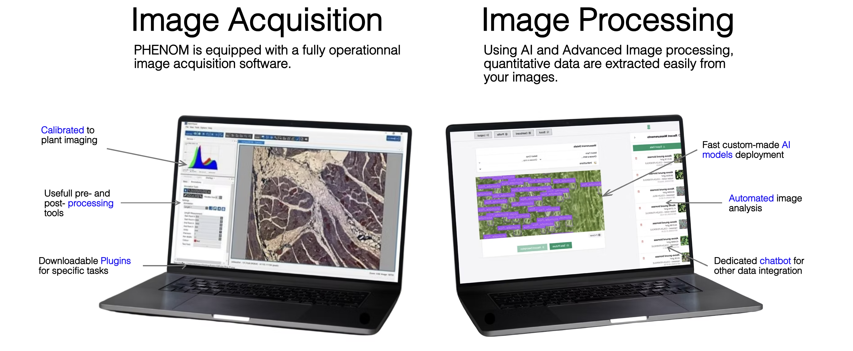
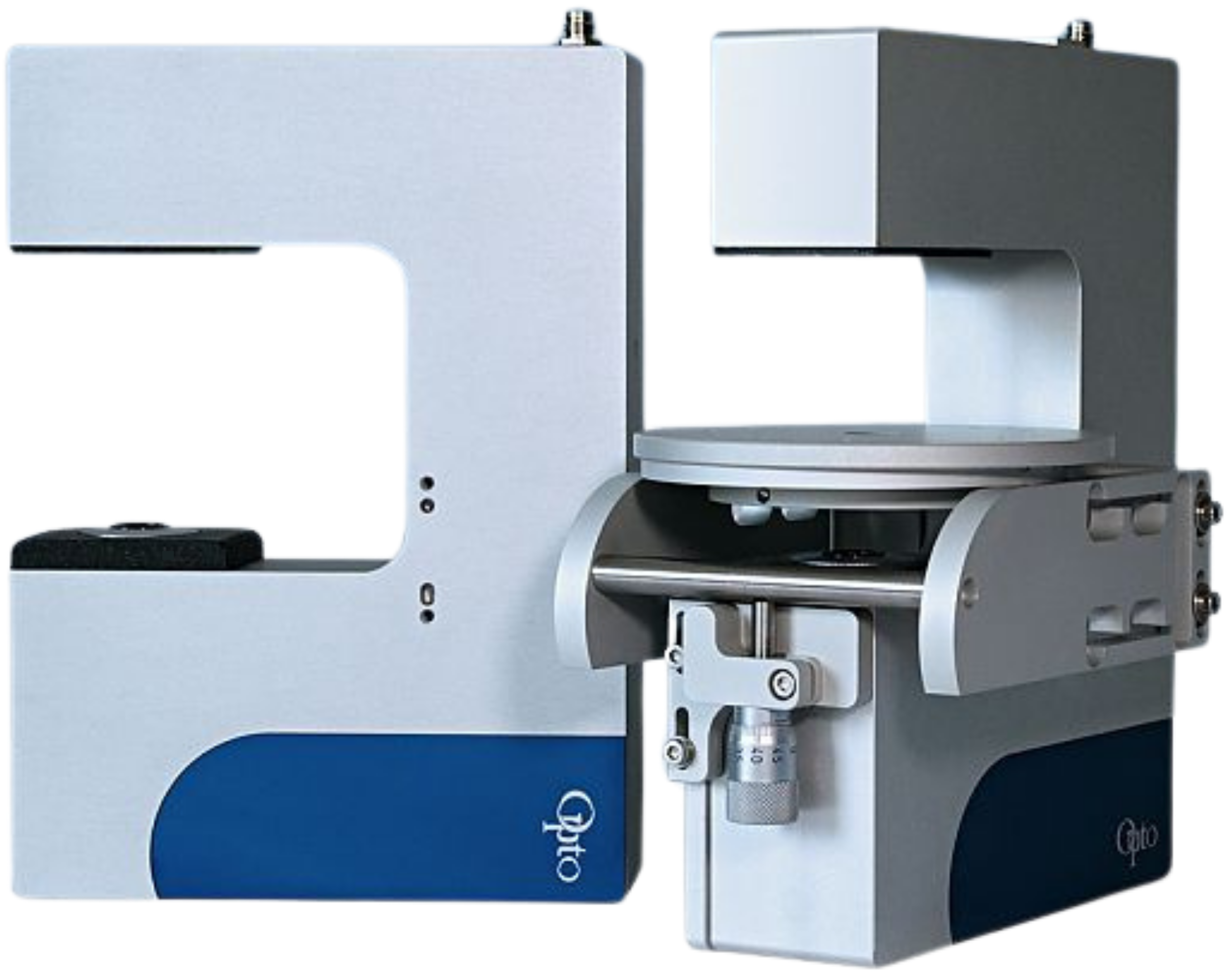
PHENOM is MIAtecs' bench-top microscopy platform for continuous, non-destructive stomatal imaging — from 15 minutes setup to 24h time-lapse experiments, powered by AI analysis.
Stomata imaged directly on an intact parsley leaf, without any preparation or cutting. The leaf is placed on the PHENOM stage and the first image is acquired within 15 minutes. Open and closed stomata are detected and annotated automatically in real time.
A tomato leaf is imaged over a continuous time-lapse experiment under controlled water stress. Individual stomata are tracked and their aperture measured at each time point — opening and closing transitions are captured in real time, providing a precise kinetics profile of stomatal response to stress.
Using PHENOM on Lilium asiatica, MIAtecs achieved sub-cellular resolution imaging of guard cells in vivo — revealing previously undescribed stomatal dynamics with unprecedented clarity. This level of precision opens new avenues for fundamental research in stomatal physiology and plant water relations.
A banana leaf is imaged over a 15-minute water-stress kinetics experiment. Individual stomata (#1, #2, #3) are tracked and their aperture measured at each time point — open/closed transitions captured in real time using MIAtecs AI models.
Whether you need short-term access or a long-term setup, let's discuss what works for your experiment.
Get in touch